


Veterinary Tech Toolbox: Mastering the SonoEye P5 VET – Tips & Tricks for Efficient Ultrasound Diagnostics
The SonoEye P5 VET is revolutionizing veterinary practice with its portable, high-definition color Doppler ultrasound capabilities. Whether you're in a clinic, farm, or emergency field setting, this handheld device offers real-time imaging, exceptional mobility, and user-friendly operation.
But how can veterinarians maximize its potential? In this guide, we’ll explore practical tips, workflow optimizations, and expert insights to help you get the most out of your SonoEye P5 VET.
1. Getting Started: Key Features & Setup
Why SonoEye P5 VET Stands Out
-
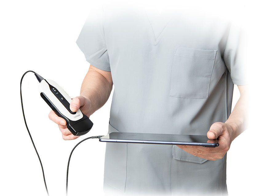
Ultra-portable design – Weighing just 420g, it fits in your pocket yet delivers high-resolution imaging.
-
Real-time color Doppler – Essential for cardiac, vascular, and abdominal assessments.
-
Multi-species compatibility – Works for small animals (dogs, cats), exotics, and large animals (horses, cattle).
-
Wireless connectivity – Stream images to tablets or PCs for remote consultations & teaching.
First-Time Setup Tips
✔ Charge fully before first use – Ensure uninterrupted sessions.
✔ Install the latest software – Check for firmware updates via the manufacturer’s app.
✔ Calibrate the probe – Run a quick system check to optimize image clarity.
2. Optimizing Your Ultrasound Workflow
Probe Selection & Handling
-
Use the linear probe (7.5-10MHz) for superficial structures (thyroid, tendons, small organs).
-
The microconvex probe (3.5-5MHz) is ideal for abdominal & cardiac scans in medium to large animals.
-
Apply adequate gel – Unlike bulky machines, the SonoEye’s compact design requires a thin, even layer for best contact.
Shortcut Keys & Touchscreen Efficiency
-
Quick freeze/measure – Use the side buttons to capture images instantly without disrupting flow.
-
Preset modes – Save customized settings for common scans (e.g., bladder, heart, pregnancy).
-
Gesture zoom – Pinch-to-zoom on the touchscreen for detailed lesion evaluation.
3. Advanced Applications: Doppler & Emergency Use
Mastering Color Doppler for Vascular Exams
-
Adjust the Doppler gain to avoid noise while maintaining sensitivity.
-
Angle correction is critical – Keep the probe at <60° for accurate blood flow readings.
-
Use pulsed-wave (PW) Doppler for quantitative measurements (e.g., renal artery stenosis in cats).
Emergency & Point-of-Care Scenarios
-
FAST exams (Focused Assessment with Sonography for Trauma) – Quickly detect free fluid in abdomen/thorax.
-
Guided cystocentesis – Improve accuracy while reducing patient stress.
-
Field triage – Assess colic in horses or bloat in cattle without moving the animal.