Pet Ultrasound Exams Demystified: Understanding Your SonoEye Color Doppler Report
Ultrasound examinations have become an essential tool in veterinary medicine, offering a non-invasive way to diagnose and monitor various health conditions in pets. However, for many pet owners, reading an ultrasound report can feel like deciphering a complex code.
In this blog post, we’ll break down the key components of a SonoEye Color Doppler Ultrasound Report, helping you understand what those images and terms mean for your furry friend’s health.

1. What Is a SonoEye Color Doppler Ultrasound?
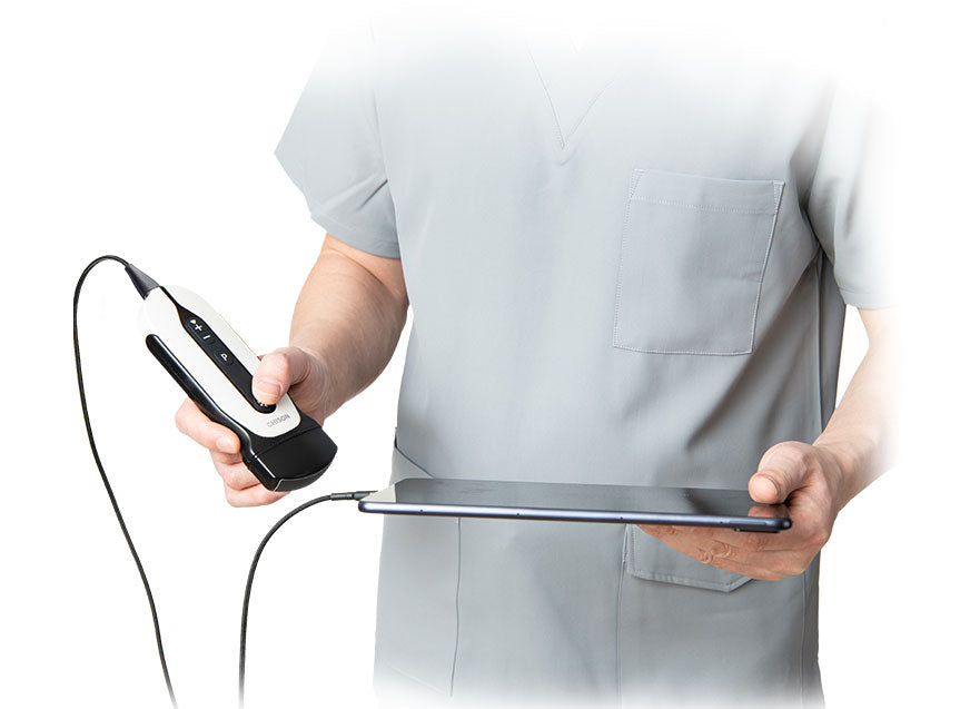
A SonoEye ultrasound is a portable, high-resolution imaging device that uses sound waves to create real-time images of your pet’s internal organs. The Color Doppler feature adds another layer of detail by visualizing blood flow, helping veterinarians detect abnormalities like blockages, tumors, or heart defects.
2. Key Sections of the Ultrasound Report
A. Patient & Clinical History
This section includes:
-
Pet’s name, age, breed, and weight
-
Reason for the scan (e.g., abdominal pain, heart murmur, pregnancy check)
-
Previous medical conditions that might affect interpretation
B. Imaging Findings
Here, the veterinarian describes what they see in the ultrasound images. Common terms include:
-
Echogenicity (how tissues appear on ultrasound):
-
Hyperechoic (bright/white – e.g., bones, stones)
-
Hypoechoic (dark/gray – e.g., fluid, some tumors)
-
Anechoic (black – e.g., urine, cysts)
-
-
Organ Measurements (liver, kidneys, heart chambers) – compared to normal ranges
-
Texture & Margins (smooth, irregular, or nodular)
C. Color Doppler Findings
This part assesses blood flow:
-
Direction & Speed: Normal flow (red/blue) vs. turbulence (mixed colors)
-
Stenosis (narrowing of vessels)
-
Shunts (abnormal blood pathways)
D. Diagnosis & Recommendations
Based on the findings, the vet may suggest:
-
Further tests (blood work, X-rays, biopsy)
-
Treatment options (medication, surgery, diet change)
-
Follow-up scans to monitor progression
3. Common Conditions Detected by Ultrasound
-
Abdominal Issues: Liver disease, bladder stones, intestinal blockages
-
Cardiac Problems: Heart valve defects, enlarged chambers
-
Reproductive Health: Pregnancy monitoring, pyometra (uterine infection)
-
Tumors & Cysts: Location, size, and vascularity (blood supply)
4. Why Choose SonoEye?
-
Portable & Fast: Ideal for emergency cases and in-clinic use.
-
High-Resolution Imaging: Clearer pictures for accurate diagnosis.
-
Real-Time Analysis: Immediate results for quicker treatment decisions.
5. Final Tips for Pet Owners
✔ Ask Questions – If a term is unclear, ask your vet to explain.
✔ Compare Follow-Ups – Track changes over time if your pet has chronic issues.
✔ Stay Calm – Not all abnormalities are serious; some just need monitoring.
Understanding your pet’s SonoEye ultrasound report empowers you to make informed decisions about their care. With this guide, you’re one step closer to decoding the mysteries of veterinary imaging!


